


Nuclear Medicine & Theranostics
PET-CT diagnostics and targeted radionuclide therapies such as Lutetium-177 PSMA and radioiodine.
Quick answer
Nuclear Medicine is a medical unit that uses small amounts of radioactive substances and specialized imaging to diagnose, stage, and monitor many conditions, and in some cases to deliver targeted treatment. At Acibadem in Turkey, Nuclear Medicine specialists perform evaluations such as PET/CT, SPECT/CT, and scintigraphy and work with other departments to plan patient-specific care.
What our nuclear medicine unit covers — and who it is for
Nuclear medicine is the specialty that images what the body is doing rather than what it looks like. A tiny amount of a radioactive tracer is given, the body handles it the way it handles the substance it is attached to, and a camera records where it goes. The picture that results is a map of function — blood flow, metabolism, bone turnover, hormone production, drainage — and it frequently shows a problem before anything has changed shape.
That is the whole distinction, and it is worth holding on to: an MRI or a CT can show that a bone looks normal while a bone scan shows that part of it is repairing furiously. Neither is better. They answer different questions.
At Acıbadem International the work falls into four strands.
- PET imaging — metabolic and receptor-targeted imaging, used most heavily in cancer staging and response assessment.
- Conventional scintigraphy — the bone, thyroid, biliary, renal, gastric and cardiac studies that answer specific functional questions no other test answers as directly.
- Radionuclide therapy — treatment rather than imaging, where a radioactive substance is delivered to the tissue that takes it up and treats it from inside.
- Theranostics — the pairing of the two: image with a tracer to prove the target is there, then treat with the same molecule carrying a therapeutic isotope.
Where the borders sit. CT, MRI and ultrasound, and the interventional work done through a catheter, belong with radiology, which also covers how to read an imaging report in general. The nuclear stress test as a cardiac investigation — who needs one, what the result means for treatment — belongs with cardiology, which covers it in full; this unit performs and reports the imaging. Chemotherapy and systemic treatment belong with medical oncology, external beam radiotherapy with radiation oncology, and thyroid surgery with general surgery. What this unit owns is the tracer, the scan, the report and the radionuclide treatment.
Function rather than structure: why the scan is different
Three properties follow from imaging function, and each explains something patients find confusing.
It can be positive before anything looks abnormal. Bone turnover, glucose uptake and receptor expression change before size does. This is why a bone scan finds a metastasis that a radiograph shows as normal bone, and why a PET scan can restage a cancer that looks unchanged on CT.
It can also be positive when nothing is wrong. Infection, inflammation, healing fractures, recent surgery, arthritis and brown fat all take up tracer, sometimes avidly. A hot spot means the tissue is metabolically active, not that it is malignant — and the commonest error in reading these scans, by patients and by clinicians outside the specialty, is treating those two statements as the same.
The preparation is part of the test. Almost every nuclear medicine study has a preparation requirement, and unlike most imaging, ignoring it does not degrade the image — it invalidates the result. A PET scan performed on a patient whose blood glucose is high, a thyroid scan done after a CT with contrast, a gastric emptying study eaten differently from the protocol: each produces a picture that looks fine and means nothing. Preparation therefore has a section of its own.
The tracer, the dose, and the radiation question
This is the first question almost everyone asks, so it belongs near the front rather than in a footnote.
A radiotracer is an ordinary biological molecule — a sugar, a phosphate, a hormone precursor, a protein fragment — with a radioactive atom attached. The amount of the molecule itself is minute, far below any pharmacological effect, which is why tracers do not cause the reactions that iodinated CT contrast or gadolinium can. Allergic reactions to diagnostic radiotracers are very rare.
The radioactivity is chosen to be the smallest amount that gives a diagnostic image, and the isotopes used decay quickly — technetium-99m, the workhorse of conventional scintigraphy, has a half-life of about six hours, and the fluorine-18 used in PET about two hours. By the following day, very little activity remains, and what is left leaves mostly in urine.
Exposure from a diagnostic nuclear medicine study is comparable in scale to other medical imaging that uses ionising radiation, and it is justified the same way: against the question being asked and the harm of not answering it. The honest framing is that these are not zero-radiation tests and are not treated as casual ones — a scan is performed because the answer changes something. Where an alternative without radiation would answer the same question, that alternative is used.
Two practical points follow. Drinking well and passing urine after a scan clears residual tracer faster. And a scan is not a reason to avoid other people generally — the specific precautions that do apply belong to radionuclide therapy rather than to diagnostic imaging, and they are set out there.
PET-CT: what it shows and what it does not
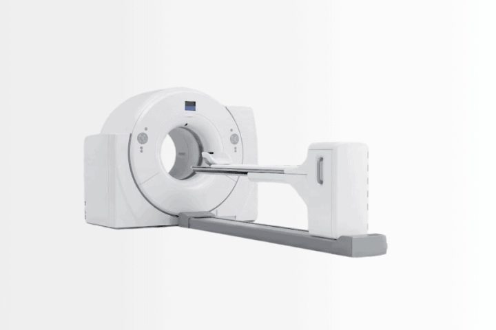
A PET CT scan combines two things in one session. The PET component maps where a tracer is concentrating; the CT component supplies the anatomy so that the finding can be located precisely. Fused, they answer a question neither answers alone: what is metabolically abnormal, and exactly where is it.
The standard tracer is FDG, a glucose analogue. Cells that consume glucose fast take up more of it, which is true of most cancers and equally true of infection, inflammation and healing tissue.
What it is used for
Staging a newly diagnosed cancer, because it examines the whole body in one pass and finds disease outside the expected field. Assessing response, since metabolic activity falls before a mass shrinks and sometimes when it never shrinks at all. Detecting recurrence when a tumour marker rises but conventional imaging looks normal. Planning radiotherapy, where the metabolically active volume rather than the visible mass defines the target. And finding a primary tumour when a metastasis has appeared and its origin is unknown.
Where it is weak
Small lesions below the resolution of the scanner are missed regardless of how active they are. Some tumours are not FDG-avid — several well-differentiated and slow-growing cancers among them — and a negative PET in those does not exclude disease. The brain uses glucose constantly, so brain metastases are poorly assessed by FDG and are imaged by MRI instead. And the false positives listed above are common enough that a solitary hot spot is confirmed rather than acted on.
The number quoted on the report, the SUV, expresses how concentrated the tracer is relative to a body-wide average. It is useful for comparing the same lesion on successive scans in the same patient on the same scanner. It is not a malignancy score, and there is no threshold above which something is cancer — a point worth knowing before reading one’s own report.
PET MRI combines the same functional imaging with MRI instead of CT, offering better soft-tissue contrast and less radiation, and it is used where those advantages matter, particularly in the pelvis, the liver and in children.
PSMA and DOTATATE: the targeted PET scans
These two changed practice in their fields, and they work on a different principle from FDG: instead of measuring how much sugar a cell consumes, they bind to a protein the tumour displays on its surface.
PSMA PET targets prostate-specific membrane antigen, expressed strongly by most prostate cancers. It detects disease at PSA levels where conventional bone scans and CT show nothing, which matters most in two situations: staging before treatment where the plan depends on whether disease is confined, and finding where recurrence sits when PSA rises after treatment. Its sensitivity is also its trap — it finds small-volume disease that older imaging never saw, and what to do about a finding that would once have been invisible is a decision for the multidisciplinary team rather than an automatic escalation.
A DOTATATE scan targets somatostatin receptors, which neuroendocrine tumours express densely. It has largely replaced the older octreotide scan, locates primary tumours that other imaging misses, and does something further: because it demonstrates that the receptor is present, it identifies patients who can be treated with the matching therapy described under theranostics.
Both are examples of the principle that defines the modern specialty — the scan does not merely describe the disease, it selects the treatment.
The bone scan
A bone scan maps bone turnover. The tracer binds where bone is actively remodelling, so anything that makes bone repair — a metastasis, a fracture, infection, arthritis, a healing surgical site — appears as a hot spot.
Its main uses are searching the whole skeleton for metastatic disease, finding a stress fracture that radiographs do not show, investigating unexplained bone pain, assessing whether a joint replacement has loosened or become infected, and identifying complex regional pain syndrome.
Two things about the result matter. The scan is extremely sensitive and correspondingly non-specific: degenerative change in the spine and old rib fractures light up routinely, and interpreting the pattern is what separates them from disease. And after successful treatment of a bone metastasis, the scan can appear worse before it appears better, because healing bone is metabolically active — the flare phenomenon, and a genuine cause of unnecessary alarm at the first follow-up.
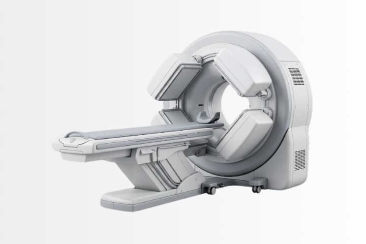
A SPECT-CT, which adds cross-sectional detail to the scintigraphy, is frequently added precisely to tell an arthritic facet joint from a vertebral metastasis.
HIDA scan: imaging the gallbladder and bile ducts
A HIDA scan — hepatobiliary scintigraphy — follows a tracer taken up by the liver and excreted into bile, so it shows whether bile is actually flowing. It answers questions ultrasound cannot, and it is the study most often requested when the ultrasound was normal and the symptoms were not.
What it answers
In suspected acute cholecystitis, failure of the gallbladder to fill indicates that the cystic duct is obstructed — the direct finding, rather than the indirect signs seen on ultrasound. In suspected biliary obstruction, it shows whether bile reaches the bowel. After gallbladder or bile duct surgery it detects a bile leak. And it establishes whether a biliary-enteric surgical connection is draining.
The gallbladder ejection fraction
The commonest reason a HIDA scan is requested in someone whose ultrasound found no stones is suspected functional gallbladder disorder — right upper abdominal pain, typical in character, with a gallbladder that looks normal. A drug that makes the gallbladder contract is given during the scan and the proportion of bile expelled is measured, giving an ejection fraction.
Two honest qualifications belong with that number. A low ejection fraction supports the diagnosis but does not by itself establish that removing the gallbladder will relieve the symptoms, and the strength of that link is genuinely debated. And the drug reproduces the patient’s pain in some people, which is informative rather than a complication. The decision that follows the result belongs with gastroenterology and the surgical team, not with the scan.
Gastric emptying study
A gastric emptying study measures how fast the stomach empties, and it is the reference test for gastroparesis. A standardised meal is labelled with a tracer and images are taken over several hours, giving the percentage of the meal still in the stomach at defined times.
It is used for nausea, vomiting, early fullness, bloating and unexplained poor diabetic control, and it also detects the opposite problem — rapid emptying, which follows some gastric and bariatric surgery and produces its own symptoms.
The preparation is the whole test and is unusually strict, because the result is a number compared against a standard protocol. The meal must be the protocol meal rather than a substitute. Medicines that speed or slow the stomach are stopped beforehand under the instruction of the doctor who prescribed them. Blood glucose must be reasonably controlled on the day, because a high glucose slows emptying by itself and produces a falsely abnormal result. Smoking on the morning of the test alters the result. And the study runs for several hours rather than minutes, which surprises people who have arranged their day around a scan.
Thyroid scan and uptake
A thyroid scan shows which parts of the thyroid are actively taking up iodine, and the thyroid uptake scan measures how much. Together they answer a question blood tests cannot: not whether the thyroid is overactive, but why.
That distinction determines treatment. An overactive gland taking up avidly and uniformly indicates Graves’ disease. One or more discrete areas taking up while the rest is suppressed indicates a toxic nodule or a toxic multinodular goitre. Low uptake in a thyrotoxic patient points to thyroiditis — inflammation releasing stored hormone rather than a gland making too much — which is treated entirely differently and never with radioactive iodine.
Nodules are described as hot or cold according to whether they take up more or less than surrounding tissue. Cold nodules are far commoner and the great majority are benign; the finding directs whether a nodule is sampled rather than deciding anything by itself, and the sampling and its interpretation belong with pathology.
One preparation point causes more cancelled thyroid studies than any other: recent iodinated contrast from a CT scan floods the body with iodine and blocks uptake for weeks. Amiodarone does the same for months. Some medicines and supplements interfere too. Anyone who has had a contrast CT recently should say so before the appointment rather than on arrival.
Radioactive iodine treatment
Radioactive iodine is the specialty’s oldest targeted therapy and remains one of medicine’s cleanest examples of the principle: the thyroid is the only tissue that concentrates iodine, so iodine carrying a therapeutic isotope treats the thyroid and largely spares everything else.
It is used for two different purposes that are worth separating. In hyperthyroidism it reduces the amount of functioning thyroid tissue, as an alternative to long-term medication or surgery. In differentiated thyroid cancer it is given after surgery to destroy remaining thyroid tissue and any residual disease.
The radioactive iodine side effects worth knowing about are mostly short-lived: a sore or swollen neck for a few days as the treated tissue reacts, a dry or altered taste, dry mouth from temporary salivary gland irritation, and nausea. The expected outcome in hyperthyroidism is a different matter and is worth stating plainly rather than presenting as a complication: many patients become hypothyroid afterwards and need lifelong thyroid hormone replacement. That is frequently the intended endpoint rather than a failure, because a reliably underactive thyroid corrected with a daily tablet is easier to live with than a fluctuating overactive one.
Treatment doses require preparation — a low iodine diet for a period beforehand so the thyroid is iodine-hungry when the dose arrives, and adjustment of thyroid medication, both directed by the treating team. Pregnancy must be excluded, breastfeeding must have stopped well beforehand, and conception is deferred for a defined interval afterwards. The precautions around other people after a therapeutic dose are real and are set out below.
Which patients should have radioactive iodine rather than surgery or medication is a decision made with endocrinology.
Parathyroid scan
A parathyroid scan, usually a sestamibi study, exists to answer one question: when blood tests show a parathyroid gland is overactive, which of the four is it?
That question matters because the answer changes the operation. A confidently localised single adenoma allows a small targeted incision rather than a bilateral neck exploration — shorter surgery, less dissection, faster recovery. A sestamibi scan works because the abnormal gland retains the tracer longer than normal tissue does, so delayed images show it while the thyroid has washed out. SPECT-CT is routinely added to give the surgeon a three-dimensional position rather than a flat one.
Two honest limits. Small glands, multiple gland disease and hyperplasia localise less reliably, and a negative scan does not mean the biochemistry was wrong — it means the operation will be planned differently. And the scan localises rather than diagnoses: the diagnosis of hyperparathyroidism is biochemical, made before any scan is ordered.
Myocardial perfusion imaging
Myocardial perfusion imaging compares blood flow to the heart muscle at rest and under stress, either from exercise or from a drug that dilates the coronary circulation. A region that receives enough blood at rest but not under stress indicates a significant narrowing; a region that receives too little in both indicates muscle that has already been damaged.
What makes it useful is that it measures the physiological consequence rather than the anatomy. A narrowing seen on an angiogram is not always the narrowing causing symptoms, and this study helps establish which lesion actually matters — which is why it is used to decide whether an intervention is warranted, and to assess whether damaged muscle would recover if blood flow were restored.
Preparation is specific and frequently missed: caffeine blocks the drugs used for pharmacological stress, so coffee, tea, chocolate and cola are avoided for a defined period beforehand, and certain cardiac medicines are held under the treating cardiologist’s instruction.
The clinical use of the result — who needs the test, what a positive result means for treatment, and how it sits alongside CT coronary angiography — belongs with cardiology, which covers the nuclear stress test in full.
DaTscan and other brain studies
A DaTscan images the dopamine transporter in the brain, showing whether the dopamine-producing nerve terminals of the basal ganglia are intact. It is used for one specific and genuinely difficult question: distinguishing a parkinsonian syndrome caused by loss of those neurons from conditions that look similar but leave them intact — essential tremor, drug-induced parkinsonism and some vascular presentations.
Its limitation is important and frequently misunderstood. An abnormal DaTscan confirms that dopaminergic neurons have been lost; it does not distinguish Parkinson’s disease from the other conditions that also lose them, such as multiple system atrophy and progressive supranuclear palsy. It answers whether, not which, and the diagnosis remains clinical, made by neurology.
Two further brain studies belong here. FDG PET of the brain shows patterns of reduced metabolism that help distinguish dementia subtypes, and amyloid or tau PET demonstrates the presence of the corresponding protein — increasingly relevant now that treatments requiring proof of amyloid exist.
Renal scan and lymphoscintigraphy
Renal scan
A renal scan measures what each kidney contributes and whether urine drains freely, which ultrasound cannot do. Its two central uses are separating obstruction from a dilated but unobstructed system — the question that decides whether a child with antenatal hydronephrosis needs an operation — and measuring split function before a kidney is removed or a donor is accepted. A DMSA study images cortical scarring instead, which is how the damage from childhood urinary infection and reflux is assessed. The clinical management belongs with nephrology, urology and paediatric surgery.
Lymphoscintigraphy
Lymphoscintigraphy maps lymphatic drainage. Its commonest use is sentinel lymph node mapping: a tracer injected around a tumour travels to the first node draining it, which the surgeon then finds and removes, so that a full nodal clearance and its long-term consequences can be avoided when that node is clear. It is standard in breast cancer and melanoma. The same technique also images lymphoedema, showing where drainage has failed and whether surgical lymphatic reconstruction is feasible.
Imaging infection and inflammation
When infection is suspected but nobody can find it, this specialty has two tools that look for it functionally rather than structurally.
A gallium scan accumulates in infection, inflammation and some tumours, and it retains a role in chronic infection, spinal infection and sarcoidosis. A labelled white cell scan is more specific for bacterial infection: the patient’s own white cells are labelled and reinjected, and they travel to where they are being recruited. It is particularly useful for infected joint prostheses and diabetic foot infection, where distinguishing infected bone from the changes of neuropathic arthropathy is genuinely hard.
FDG PET has taken over much of this work, especially for fever of unknown origin, large vessel vasculitis and infected vascular grafts, because it examines the whole body at once. The clinical side — which organism, which antimicrobial, how long — belongs with infectious diseases.
Theranostics: seeing the target, then treating it
Theranostics is the idea that gives this specialty its current momentum, and it is simpler than the word suggests. The same molecule that carries an imaging isotope to a target can carry a therapeutic isotope instead. The scan proves the target is present in that particular patient, and the treatment then goes to the same place.
This inverts the usual order. Rather than giving a treatment and waiting to see whether it works, the imaging step establishes in advance that the tumour expresses what the treatment binds to — and a patient whose scan shows the target is absent is spared a treatment that could not have worked.
Two pairings are established in routine practice. In prostate cancer, PSMA PET identifies patients whose disease expresses PSMA, and lutetium 177 PSMA therapy then delivers radiation to those same sites. In neuroendocrine tumours, a DOTATATE scan demonstrates somatostatin receptor expression and lutetium-177 DOTATATE therapy treats accordingly. MIBG follows the same logic in neuroblastoma and phaeochromocytoma: an MIBG scan demonstrates uptake first, and MIBG therapy then treats the sites that took it up.
These are given over several cycles with blood counts, kidney function and salivary function monitored, and they are decided at a multidisciplinary meeting with medical oncology rather than requested directly. They are not first-line treatments and they are not cures; what they offer, in selected patients, is disease control with a side effect profile that differs from chemotherapy.
Radioembolization works on a related principle for liver tumours — Y 90 microspheres, yttrium-90, delivered into the artery feeding the tumour — and is performed jointly with interventional radiology, which places the catheter.
Preparing for a scan, and why it decides the result
More nuclear medicine studies are wasted by preparation than by anything technical, and the reason is structural: the tracer is handled by normal physiology, so anything that alters that physiology alters the picture.
- PET-CT. Fasting for a defined period, because insulin drives glucose and its tracer into muscle instead of tumour. Blood glucose is checked on arrival and a high level may mean rescheduling. No strenuous exercise for a day beforehand — exercised muscle takes up avidly. Keeping warm before and during the uptake period matters, because cold activates brown fat, which produces striking uptake in the neck and chest and can obscure genuine disease.
- Thyroid studies. Recent iodinated CT contrast blocks uptake for weeks and amiodarone for months. Iodine-rich supplements and certain medicines interfere. Declare a recent contrast CT before the appointment, not on the day.
- Cardiac perfusion. No caffeine for a defined period, because it blocks the pharmacological stress agents. Some cardiac medicines are held, on the instruction of the prescribing doctor.
- Gastric emptying. The protocol meal exactly, controlled blood glucose, no smoking that morning, and prokinetic or slowing medicines stopped beforehand as directed.
- HIDA. Fasting for a defined period so the gallbladder is not already contracted, and opioids avoided beforehand because they contract the sphincter of Oddi.
- Everything. Pregnancy is asked about every time and is asked directly rather than assumed. Breastfeeding requires specific advice per tracer and is almost never a reason to cancel — it is a reason to plan. Bring previous imaging and reports, because comparison changes interpretation here as much as anywhere.
The exact intervals, doses and which medicines to hold are given by the treating team for the individual study, because they differ by tracer, by protocol and by patient.
Radiation, pregnancy, and being near other people
Diagnostic scans and radionuclide therapy are different situations and conflating them causes unnecessary fear.
After a diagnostic scan, the activity is small and short-lived. Ordinary contact with adults is not restricted. A common-sense precaution is to limit prolonged close contact with pregnant women and young children for the remainder of the day, and drinking well and passing urine clears the residual tracer faster. Travellers should know that radiation portal monitors at airports and borders are sensitive enough to detect a recent scan for days afterwards, which is why a letter stating the date, the tracer and the dose is worth carrying.
After radionuclide therapy the precautions are genuine and specific: a period of distance from others, particularly pregnant women, infants and young children, separate sleeping arrangements for a defined time, careful bathroom hygiene, and separate laundering. Higher-dose treatments require a period in a dedicated room. The exact durations depend on the isotope, the dose and the household, and they are given individually by the treating team rather than as a general rule.
Pregnancy is a contraindication to radionuclide therapy and is excluded before treatment. Conception is deferred for a defined interval afterwards, for both women and men. Breastfeeding must have stopped well before therapy, and for diagnostic scans it is usually a matter of a defined interruption rather than stopping.
A final point worth saying plainly: after a diagnostic scan a person is not radioactive in any way that endangers their family, and after therapy the precautions exist to keep exposure to others as low as reasonably achievable rather than because they are known to be dangerous at those levels.
What nuclear medicine cannot do
It cannot tell malignant from inflammatory uptake on its own. Infection, healing and arthritis are metabolically active, and a hot spot is a finding to be explained rather than a diagnosis.
It cannot see below its resolution. Small deposits are missed regardless of how avid they would have been, which is why a negative scan is read alongside the clinical picture rather than as an all-clear.
It cannot exclude disease that does not take up the tracer. Several slow-growing tumours are not FDG-avid, and a prostate cancer that does not express PSMA will not appear on a PSMA PET however extensive it is.
It cannot give a number that means cancer. The SUV compares one measurement with another in the same patient; there is no threshold above which a finding is malignant.
It cannot overcome preparation. A PET performed with a high blood glucose, a thyroid scan after contrast, a cardiac study after caffeine — each returns an image that looks convincing and answers nothing.
It cannot make a therapy work where the target is absent. That is the point of the imaging step in theranostics, and a scan showing no expression is a useful result rather than a failed one.
Your multidisciplinary team
The nuclear medicine physician decides which study answers the question, supervises the tracer administration, interprets the images and delivers the radionuclide therapies. The medical physicist calculates and verifies doses, manages scanner quality control and handles the dosimetry that makes therapy individual rather than standard. The radiopharmacist prepares the tracers, several of which are made or labelled shortly before use because they decay so quickly. The nuclear medicine technologist positions the patient, acquires the images and applies the preparation protocol that determines whether the study is valid. The radiation safety officer manages the therapy rooms, the waste and the precautions given to families.
Around them: medical oncology at every tumour board where staging and response are decided, radiology for the cross-sectional imaging that is read alongside and for the catheter work in radioembolization, radiation oncology where PET defines the treatment volume, cardiology for perfusion imaging, endocrinology for thyroid and parathyroid disease, urology for prostate cancer, neurology for movement disorders and dementia, gastroenterology for biliary and motility studies, pathology wherever a scan finding is sampled, infectious diseases for infection imaging, and breast health for sentinel node mapping.
The international patient journey
Three patterns cover almost everything, and the preparation differs sharply between them.
A scan that is not available locally. PSMA PET, DOTATATE PET, PET-MRI and specialised SPECT-CT are the commonest reasons. These are arranged as a short visit, but the preparation begins before departure: the fasting or dietary requirement, the medicines to hold, and above all any recent iodinated contrast, which can make a thyroid study impossible for weeks. Sending the clinical question and the previous imaging in advance is what determines that the right study is booked.
Radionuclide therapy. Lutetium-177 PSMA or DOTATATE, or radioactive iodine for thyroid disease — the latter one of the three Graves’ treatment doors endocrinology weighs with you. These are planned rather than booked. Eligibility is established first, usually from a scan demonstrating the target, and the assessment happens before travel. Treatment runs in cycles with intervals between them, and the isolation and travel restrictions afterwards are part of the plan rather than an afterthought — including the interval before flying, which is set by the dose.
A second reading of a scan performed elsewhere. Possible and useful, and it needs the raw imaging data rather than a report or printed images. The same caveat as in radiology applies: a re-read cannot recover what the acquisition never captured, and if the preparation was wrong, the answer will be that the study needs repeating.
Three practical notes. Bring the actual imaging on disc or through a transfer link, together with previous reports and any tumour marker results — a PSA trend or a chromogranin level changes how a scan is interpreted. Declare recent contrast CT, recent nuclear studies and current medicines before the appointment is fixed rather than on arrival. And carry a letter after any scan or therapy, because border radiation monitors will detect it for days and a letter turns a long delay into a short conversation.
Technology used in this program
Send your reports and get a plan from a tumour board, not a single doctor
Pathology, imaging and previous treatment history are enough to start. Written recommendation within 48 hours, including trial eligibility.